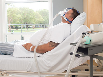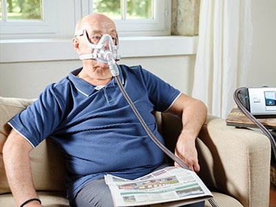
Obesity hypoventilation syndrome (OHS)
OHS is frequent in obese individuals and associated with high mortality, yet is commonly misdiagnosed.1 On this page, you will learn more about this condition, how it is diagnosed and what the available treatment options are.
PART 1
Understanding OHS
What is OHS?
Obesity hypoventilation syndrome is defined as daytime hypercapnia PaCO2>45 mm Hg and sleep-disordered breathing, occurring in an obese (BMI≥30 kg.m²) individual without other causes of hypoventilation, such as COPD or neuromuscular disease.1,2
Key symptoms
Symptoms are often non-specific and include excessive daytime sleepiness, fatigue, dyspnoea and headaches, all of which can disrupt a patient’s daily routine and impact quality of life (QoL).2,3 Many of these symptoms are also observed in patients with obstructive sleep apnoea (OSA), which affects around 90% of patients with OHS.1,2
Pathophysiology
The pathophysiology of OHS is complex and thought to be related to three major mechanisms.1,3,4
- Mechanical changes to the respiratory system lead to increased work of breathing and are due to increased adipose tissue in the abdomen and chest wall
- A diminished central respiratory drive due to a resistance to leptin, a powerful stimulant of ventilation
- Breathing abnormalities during sleep including obstructive sleep apnoea or non-obstructive hypoventilation, together with disrupted compensatory mechanisms, lead to hypercapnia
Comorbidities
Up to 88% of OHS patients suffer from hypertension1,3
Patients with OHS often suffer from other metabolic and cardiovascular comorbidities.5 These include heart failure, coronary heart disease, insulin resistance and, in up to 88% of patients, hypertension.1,3 Comorbidities are of major importance as they are associated with greater healthcare resource use and poorer patient outcomes, highlighting the need for a holistic treatment approach in OHS.1

Understanding OHS
OHS has a complex pathophysiology and is associated with frequent comorbidities and non-specific symptoms. So how common is OHS and how is it diagnosed?
PART 2
Prevalence and diagnosis of OHS
Prevalence
31% of hospitalised adults with BMI >35kg/m² have OHS2
- Worldwide, almost 10% of adults are obese1
- Obesity affects 23% of individuals across the WHO European region,6 of whom 23% may have OHS.7
- In patients referred to sleep centres for the evaluation of sleep-disordered breathing, the prevalence of OHS is 8–23%1,7
- Moreover, the prevalence of OHS among hospitalised adults with a BMI >35 kg/m2 was found to be 31%,2 highlighting the need to consider OHS in hospitalised patients with obesity/morbid obesity
Diagnosis
OHS typically presents as an acute-on-chronic exacerbation with acute respiratory acidosis, or during a consultation with sleep/pulmonary specialists.1,2
- OHS can be diagnosed in patients with a BMI >30 kg/m2 using arterial blood gases (daytime PaCO2 >45 mmHg) and polysomnography.1,2,5,8 However it should be noted that OHS is a diagnosis of exclusion and other disorders that may cause alveolar hypoventilation should be ruled out
- Serum bicarbonate levels (<27 mmol L-1or ≥27 mmol L-1) and the presence of cardiometabolic comorbidities may also serve as an indicator of OHS severity and are included in The European Respiratory Society staging system for OHS8
Misdiagnosis/Lack of diagnosis
OHS diagnosis is often delayed, typically occurring in the fifth and sixth decades of life and misdiagnosis is common.1
In one study, 8% of all patients admitted in the ICU met the diagnostic criteria for OHS but ~75% were misdiagnosed with obstructive lung disease.1 Since mortality rates for untreated OHS are important,1 it is crucial that it is diagnosed in a timely manner.

Prevalence and diagnosis of OHS
OHS appears to be common in obese individuals, yet is often misdiagnosed as obstructive lung disease. Diagnosis requires assessment of arterial blood gases and polysomnography, together with excluding other causes of alveolar hypoventilation. Next, we will review the available treatments for OHS.
PART 3
Treatment and prognosis of OHS
OHS management strategy
When selecting the most appropriate mode of PAP therapy (CPAP or NIV), clinicians should consider the patient’s “OHS phenotype” i.e., the predominant underlying mechanism of their OHS.1
CPAP treatment
CPAP is a highly effective treatment for OSA and may help improve gas exchange in a substantial group of patients by stabilising the upper airways.1 This form of respiratory support may be more appropriate for patients with a greater number of obstructive events during sleep (Figure 1).1
NIV treatment
- NIV seems more appropriate for patients with more “pure” forms of hypoventilation and with fewer obstructive events during sleep (i.e., mild to no OSA) (Figure 1)1,5
- NIV may also be more effective for patients with comorbid pulmonary hypertension as it can improve cardiac structure and function, and reduces pulmonary systolic artery pressure in patients with OHS1
- Patients without a favourable response to initial CPAP therapy despite high levels of adherence should be switched to NIV1
- Randomised studies comparing CPAP with NIV in different phenotypes of OHS are needed12
International guidelines
The most commonly used guidelines for the treatment of OHS are those by the European Respiratory Society (ERS) and the American Thoracic Society (ATS).5,8
ERS guidelines
The ERS does not categorically recommend one form of ventilation over another, but states that CPAP treatment improves AHI, oxygen saturation, hypercapnia and ventilatory response to O2 and CO2 in a majority of OHS patients, although CPAP failure is higher in OHS compared with OSA8. In addition, NIV during sleep improves hypoventilation, sleep, QoL, and survival and is superior to lifestyle counselling.8
ATS guidelines
ATS recommends PAP treatment during sleep for stable ambulatory patients with OHS:
- CPAP rather than NIV should be offered as the first-line treatment to stable ambulatory patients with OHS and coexistent severe OSA5 who represent the majority of patients
- Patients with a greater degree of initial ventilatory failure, poorer lung function, and advanced age may be less likely to respond to CPAP and should be closely monitored5
- Patients with an inadequate response to CPAP should be switched to NIV5
Other approaches
Weight loss interventions including bariatric interventions can improve OHS and OSA, as well as cardiovascular and metabolic outcomes.5,8 The ATS recommend weight loss interventions that produce sustained weight loss of 25–30%.5
Consequences of untreated OHS
Untreated OHS is associated with significant morbidity and mortality:1
- Patients are often admitted to the hospital and require respiratory or critical care Indeed, the development of acute respiratory acidosis leading to admission to an intensive care unit is one of the most common presentations of OHS leading to diagnosis2
- Patients have a significant risk of death. Observational studies have shown all-cause mortality of 24% at 1.5–2 years,1 which is reduced with adherence to PAP therapy.9
Prognosis
NIV and CPAP can improve long-term outcomes:1
PAP treatment leads to improvements in blood gases and symptoms (such as daytime sleepiness) and improves sleep-disordered breathing.
A multimodal approach incorporating lifestyle/weight management elements together with PAP therapy may be the most effective strategy to optimise outcomes in patients with OHS.4

Treatment and prognosis of OHS
Untreated OHS can lead to hypercapnia and respiratory failure and is associated with hospitalisation and increased mortality. Supportive respiratory therapy with positive airway pressure is recommended by national and international guidelines and has been shown to improve symptoms and survival, with the choice of CPAP or NIV determined by OHS phenotype.
In conclusion, a timely diagnosis of OHS together with a multimodal approach incorporating PAP therapy with lifestyle/weight management elements may be the most effective strategy to optimise outcomes in patients with OHS.
This content is intended for health professionals only.
References
- Masa JF et al. Eur Respir Rev. 2019;28:180097. https://err.ersjournals.com/content/28/151/180097.long
- Mokhlesi B. Respir Care. 2010;55:1347–1365. https://rc.rcjournal.com/content/respcare/55/10/1347.full.pdf
- Piper AJ and Grunstein RR. Am J Respir Crit Care Med. 2011;183:292–298. https://www.atsjournals.org/doi/10.1164/rccm.201008-1280CI
- Shetty S and Parthasarathy S. Curr Pulmonol Rep. 2015;4:42–55. https://www.ncbi.nlm.nih.gov/pmc/articles/PMC4444067/
- Mokhlesi B et al. Am J Respir Crit Care Med. 2019;200:e6–e24. https://doi.org/10.1164/rccm.201905-1071ST
- World Health Organization. WHO European Regional Obesity Report 2022. Available at: https://apps.who.int/iris/bitstream/handle/10665/353747/9789289057738-eng.pdf. Accessed February 14, 2023
- Delample D et al. Eur Respir J. 2022;60:3676. https://erj.ersjournals.com/content/60/suppl_66/3676
- Randerath W et al. Eur Respir J 2017; 49: 1600959. https://erj.ersjournals.com/content/49/1/1600959.long
- Soghier I et al. Ann Am Thorac Soc 2019;16(10):1295–1303. https://doi.org/10.1513/AnnalsATS.201905-380OC
- Masa JF et al. Am J Respir Crit Care Med 2015;192(1):8695. https://doi.org/10.1164/rccm.201410-1900OC
- Masa JF et al. Lancet; 2019;393(10182):1721–1732. https://doi.org/10.1016/S0140-6736(18)32978-7
- Noda JR et al. Thorax 2017;72(3)398–399. https://thorax.bmj.com/content/thoraxjnl/72/5/398.full.pdf


